FROM
GENE TO PROTEIN
The information content of DNA is in
the form of specific sequences of nucleotides.
- DNA dictates the synthesis of proteins,
which are the links between genotype and phenotype.
The symptoms of an inherited disease reflect a person's inability
to synthesize a particular enzyme.
The one gene - one enzyme hypothesis, but not all proteins
are enzymes and yet their synthesis depends on specific genes.
The one gene - one protein hypothesis but many proteins
are composed of several polypeptides, each of which has its own
gene.
Therefore, the hypothesis has been restated as the one gene
- one polypeptide hypothesis.
Transcription and translation
are the two main processes linking gene to protein
The bridge between DNA and protein
synthesis is RNA.
- RNA is chemically similar to DNA,
except that it contains ribose as its sugar and substitutes
the nitrogenous base uracil for thymine.
An RNA molecule almost always consists of a single strand.
The specific sequence of hundreds or thousands of nucleotides
in each gene carries the information for the primary structure
of a protein, the linear order of the 20 possible amino acids.
To get from DNA, written in one chemical language, to protein,
written in another, requires two major stages, transcription
and translation.
During transcription, a DNA strand
provides a template for the synthesis of a complementary RNA strand.
Fig.
17.2
- This process is used to synthesize
any type of RNA from a DNA template.
Transcription of a gene produces a messenger RNA (mRNA)
molecule.
During translation, the information
contained in the order of nucleotides in mRNA is used to determine
the amino acid sequence of a polypeptide.
- Translation occurs at ribosomes.
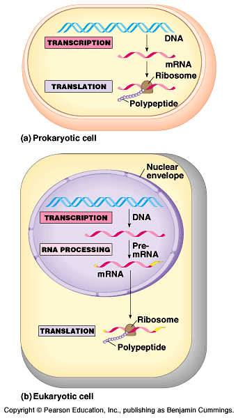
The basic mechanics of transcription and translation are similar
in eukaryotes and prokaryotes.
Because bacteria lack nuclei, transcription and translation are
coupled.
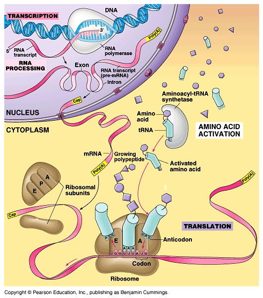
In a eukaryotic cell, almost all transcription occurs in the
nucleus and translation occurs mainly at ribosomes in the cytoplasm.
In addition, before the primary transcript can leave the
nucleus it is modified in various ways during RNA processing
before the finished mRNA is exported to the cytoplasm.
DNA -> RNA -> protein.
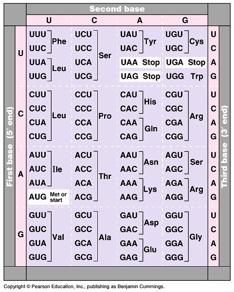
Nucleotide triplets
specify amino acids
- In the triplet code, three
consecutive bases specify an amino acid, creating 43 (64) possible
code words. Fig.
17.3.
During transcription, one DNA strand, the template strand,
provides a template for ordering the sequence of nucleotides
in an RNA transcript.
Uracil is the complementary base to adenine.
During translation, blocks of three nucleotides, codons,
are decoded into a sequence of amino acids. The codons are read
in the 5'->3' direction along the mRNA.
Each codon specifies which one of the 20 amino acids will be
incorporated at the corresponding position along a polypeptide.
Nirenberg determined the first match:
UUU coded for the amino acid phenylalanine.
- He created an artificial mRNA molecule
entirely of uracil and added it to a test tube mixture of amino
acids, ribosomes, and other components for protein synthesis.
This "poly(U)" translated into a polypeptide containing
a single amino acid, phenyalanine, in a long chain.
By the mid-1960s the entire code was
deciphered. Fig
17.4.
- 61 of 64 triplets code for amino acids.
The codon AUG not only codes for the amino acid methionine but
also indicates the start of translation.
Three codons do not indicate amino acids but signal the termination
of translation.
To extract the message from the genetic code requires specifying
the correct starting point.
This establishes the reading frame and subsequent codons
are read in groups of three nucleotides.
The genetic code must
have evolved very early in the history of life
- The genetic code is nearly universal,
shared by organisms from the simplest bacteria to the most complex
plants and animals.
In laboratory experiments, genes can be transcribed and translated
after they are transplanted from one species to another.
This has permitted bacteria to be programmed to synthesize certain
human proteins after insertion of the appropriate human genes.
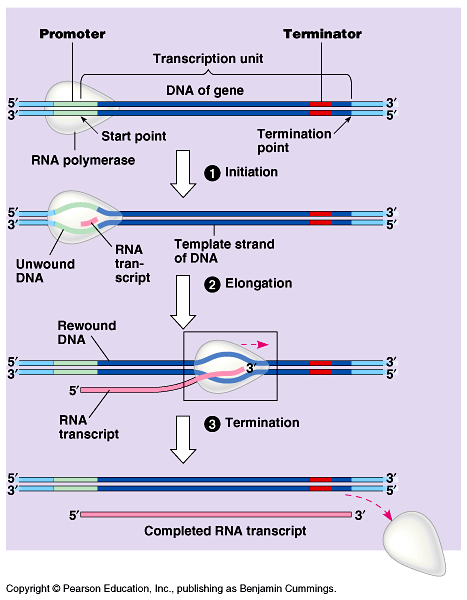
Transcription is the
DNA-directed synthesis of RNA. Fig
17.6a
- Messenger RNA is transcribed from
the template strand of a gene.
RNA polymerase separates the DNA strands and bonds the
RNA nucleotides to the 3' end of the growing polymer as they
base-pair along the DNA template.
Genes are read 3'->5', creating a 5'->3' RNA molecule.
Specific sequences of nucleotides along the DNA mark where gene
transcription begins and ends.
RNA polymerase attaches and initiates transcription at
the promotor, "upstream" of the information
contained in the gene, the transcription unit. Fig
17.7
The terminator signals the end of transcription.
Bacteria have a single type of RNA polymerase
that synthesizes all RNA molecules.
Eukaryotes have three RNA polymerases (I, II, and III) in their
nuclei.
RNA
polymerase II is used for mRNA synthesis.
Transcription can be separated into three stages:
initiation, elongation, and termination.
Initiation - The presence of a promotor sequence
determines which strand of the DNA helix is the template.
- Within the promotor is the starting
point for the transcription of a gene.
The promotor also includes a binding site for RNA polymerase
upstream of the start point.
In eukaryotes, proteins called transcription factors recognize
the promotor region, especially a TATA box, and bind to
the promotor.
After they have bound to the promotor, RNA polymerase binds to
transcription factors to create a transcription initiation
complex.
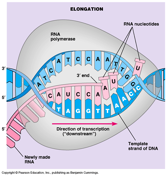
Elongation
- RNA polymerase then starts transcription. Fig
17.6b
- As RNA polymerase moves along the
DNA, it untwists the double helix, and adds nucleotides to the
3' end of the growing strand.
Behind the point of RNA synthesis, the double helix re-forms
and the RNA molecule peels away.
A single gene can be transcribed simultaneously by several RNA
polymerases at a time. This helps the cell make the encoded protein
in large amounts.
Termination -
Transcription proceeds until after the RNA polymerase transcribes
a terminator sequence in the DNA.
Transcription, the movie!
Eukaryotic cells modify RNA after
transcription
At the 5' end of the pre-mRNA molecule,
a modified form of guanine is added, the 5' cap, which
helps protect mRNA from hydrolytic enzymes. Fig
17. 8.
- At the 3' end, an enzyme adds, the
poly(A) tail.
It inhibits hydrolysis, and enables ribosome attachment and the
export of mRNA from the nucleus.
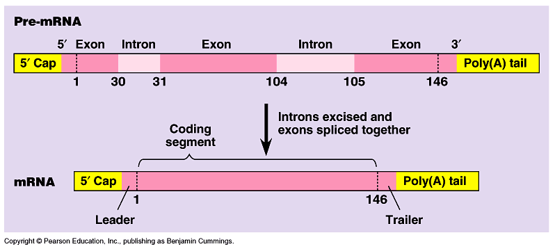
RNA splicing.
Fig
17.9
- Most eukaryotic genes and their RNA
transcripts have long noncoding stretches of nucleotides.
Noncoding segments, introns, lie between coding regions,
exons, which are translated into amino acid sequences,
plus the leader and trailer sequences.
RNA splicing removes introns and joins exons to create a mRNA
molecule with a continuous coding sequence.
This splicing is accomplished by a spliceosome. Fig
17.10.
Spliceosomes consist of a variety of proteins and several small
nuclear ribonucleoproteins (snRNPs).
Each snRNP has several protein molecules and a small nuclear
RNA molecule (snRNA).
In this process, the snRNA acts as a ribozyme, an RNA
molecule that functions as an enzyme.
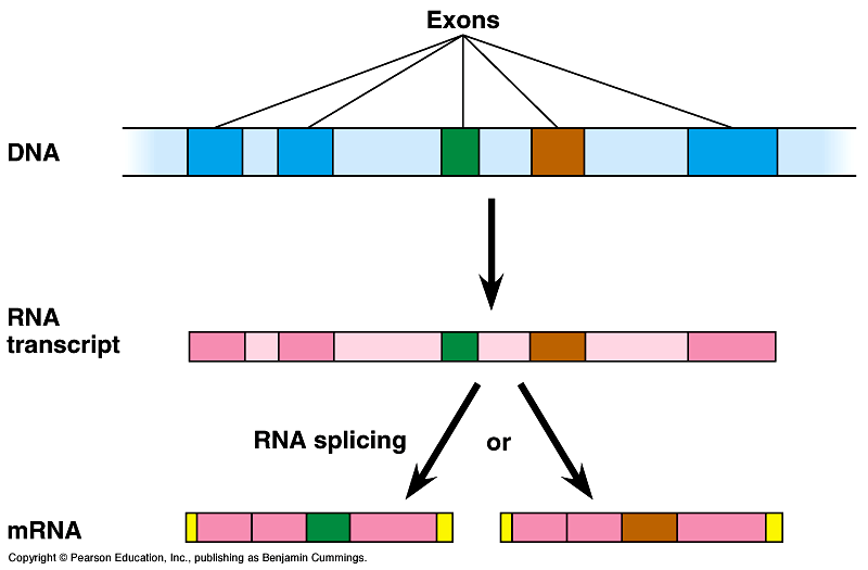
RNA splicing appears to have several functions.
1.
Some introns contain sequences that control gene activity in
some way.
2.
May regulate the passage of mRNA from the nucleus to the cytoplasm.
3.
Enables one gene to encode for more than one polypeptide.
Alternative RNA splicing gives rise to two or more different polypeptides,
depending on which segments are treated as exons. Fig
19.11.
- Proteins often have a modular architecture
with discrete structural and functional regions called domains.
Fig
17.11.
In many cases, different exons code for different domains of
a protein.
Introns increase the opportunity for recombination between two
alleles of a gene.
Exon shuffling could lead to new proteins through novel combinations
of functions.
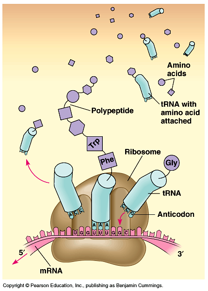
Translation is the RNA-directed
synthesis of a polypeptide. Fig
17.12.
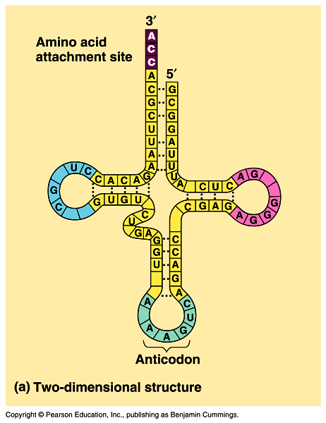
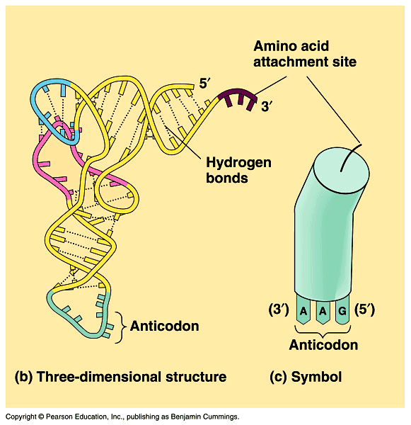
Transfer RNA
(tRNA) (2-dimensional image Fig
17.13a; 3-dimensional and symbol Fig
17.13b) transfers amino acids from the cytoplasm's pool to
a ribosome.
- The ribosome adds each amino acid
carried by tRNA to the growing end of the polypeptide chain.
During translation, each type of tRNA links a mRNA codon with
the appropriate amino acid.
Each tRNA arriving at the ribosome carries a specific amino acid
at one end and has a specific nucleotide triplet, an anticodon,
at the other.
Codon by codon, tRNAs deposit amino acids in the prescribed order
and the ribosome joins them into a polypeptide chain.
tRNA molecules are transcribed from DNA templates in the nucleus.
Each tRNA is used repeatedly.
To pick up its designated amino acid in the
cytosol.
To deposit the amino acid at the ribosome.
To
return to the cytosol to pick up another copy of that amino acid.
The anticodons of some tRNAs recognize
more than one codon. The rules for base pairing between the third
base of the codon and anticodon are relaxed (called wobble).
- At the wobble position, U on the anticodon
can bind with A or G in the third position of a codon.
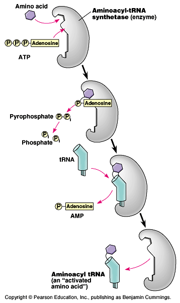
Each amino acid is joined to the correct
tRNA by aminoacyl-tRNA synthetase. Fig
17.14. The 20 different synthetases match the 20 different
amino acids.
- The synthetase catalyzes a covalent
bond between them, forming aminoacyl-tRNA or activated
amino acid.
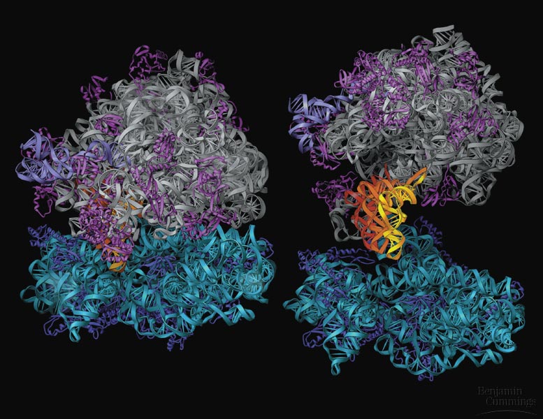
Ribosomes
facilitate the specific coupling of the tRNA anticodons with mRNA
codons.
- Each ribosome has a large and a small
subunit. Fig
17.15
These are composed of proteins and ribosomal RNA (rRNA),
the most abundant RNA in the cell.
Each ribosome has a binding site for mRNA and three binding sites
for tRNA molecules.
The P site holds the tRNA carrying the growing polypeptide
chain.
The A site carries the tRNA with the next amino acid.
Discharged tRNAs leave the ribosome at the E site.
RNA is the catalyst for peptide bond formation.
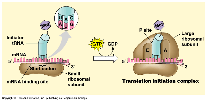
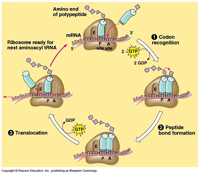
Translation can be divided into three
stages: Initiation, Elongation,
and Termination
Initiation brings together mRNA, a tRNA with the first amino
acid, and the two ribosomal subunits. Fig
17.17.
- First, a small ribosomal subunit binds
with mRNA and a special initiator tRNA, which carries methionine
and attaches to the start codon. AUG = initiator codon
Initiation factors bring in the large subunit such
that the initiator tRNA occupies the P site.
Elongation
consists of a series of three-step cycles as each amino acid is
added to the proceeding one. Fig
17.18.
- Codon recognition
Peptide bond formation
Translocation
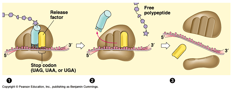
Termination
occurs when one of the three stop codons reaches the A site. Fig
17.19.
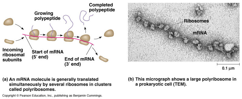
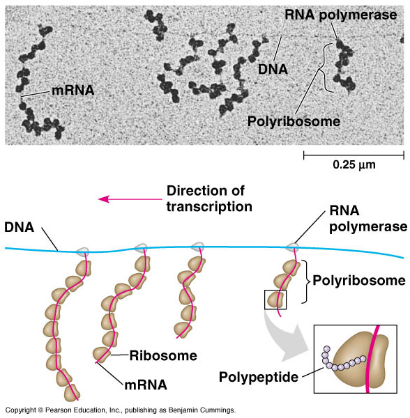
Typically a single mRNA is used to make many copies of a polypeptide
simultaneously.
Multiple ribosomes, polyribosomes, may trail along the
same mRNA. Fig
17.20.
Translation,
the movie!
During and after synthesis, a polypeptide coils and folds to its
three-dimensional shape spontaneously.
In addition, proteins may require posttranslational modifications.
- This may require additions like sugars,
lipids, or phosphate groups to amino acids.
Enzymes may remove some amino acids or cleave whole polypeptide
chains.
Two or more polypeptides may join to form a protein.
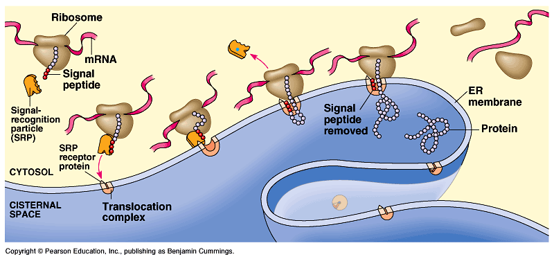
Ribosomes, free and bound.
Free ribosomes are suspended in the cytosol and synthesize
proteins that reside in the cytosol.
Bound ribosomes are attached to the cytosolic side of the
endoplasmic reticulum. Fig.
17.21.
- They synthesize proteins of the endomembrane
system as well as proteins secreted from the cell.
Translation in all ribosomes begins in the cytosol, but a polypeptide
destined for the endomembrane system or for export has a specific
signal peptide region at or near the leading end.
A signal recognition particle (SRP) binds to the
signal peptide and attaches it and its ribosome to a receptor
protein in the ER membrane.
After binding, the SRP leaves and protein synthesis resumes with
the growing polypeptide snaking across the membrane into the
cisternal space via a protein pore.
Other kinds of signal peptides are used
to target polypeptides to mitochondria, chloroplasts, the nucleus,
and other organelles that are not part of the endomembrane system.
In
these cases, translation is completed in the cytosol before the
polypeptide is imported into the organelle.
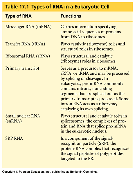
RNA plays multiple
roles in the cell: a review Table
17.1.
Comparing protein
synthesis in prokaryotes (Fig
17.22) and eukaryotes (Fig
17.25)
- One big difference is that prokaryotes
can transcribe and translate the same gene simultaneously.
The new protein quickly diffuses to its operating site.
In eukaryotes, the nuclear envelope segregates transcription
from translation.
In addition, extensive RNA processing is inserted between these
processes.
Point mutations can
affect protein structure and function
Mutations
are changes in the genetic material of a cell (or virus).
- These include large-scale mutations
in which long segments of DNA are affected (for example, translocations,
duplications, and inversions).
A chemical change in just one base pair of a gene causes a point
mutation.
If these occur in gametes or cells producing gametes, they may
be transmitted to future generations.
For example, sickle-cell disease is caused by a mutation of a
single base pair in the gene that codes for one of the polypeptides
of hemoglobin. Fig
17.23
A point mutation that results in the replacement of a pair of
complementary nucleotides with another nucleotide pair is called
a base-pair substitution. Fig
17.24a.
Some base-pair substitutions have little or no impact on protein
function.
Missense mutations are those that still code for an amino
acid but change the indicated amino acid.
Nonsense mutations change an amino acid codon into a stop
codon, nearly always leading to a nonfunctional protein.
Insertions and deletions are additions or losses
of nucleotide pairs in a gene. Fig
17.24b.
These have a disastrous effect on the resulting protein more
often than substitutions do.
Unless these mutations occur in multiples of three, they cause
a frameshift mutation.
All the nucleotides downstream of the deletion or insertion will
be improperly grouped into codons.
Mutations can occur during DNA replication, DNA repair, or DNA
recombination.
These are called spontaneous mutations.
Mutagens
are chemical or physical agents that interact with DNA to cause
mutations.
- Physical agents include high-energy
radiation like X-rays and ultraviolet light.
This makes sense because most carcinogens are mutagenic and most
mutagens are carcinogenic.
What is a gene?
- The Mendelian concept of a gene views
it as a discrete unit of inheritance that affects phenotype.
A gene is a specific nucleotide sequence along a region of a
DNA molecule.
A gene is a region of DNA whose final product is either
a polypeptide or an RNA molecule.