|

|
|
Embryonic Stem Cell Technologies |
|
|
Embryonic Stem
(ES) Cells |
I am interested in the methods of culture, lineage selection and applications of ES cells, in order to develop quantitative methods to study tissue development and to assess novel disease therapies.
Much interest has been brought to bear upon ES cells, as these cells have the unique ability to become any cell type within the body and to continue to grow. Therefore, these cells offer the opportunity to replace lost or damaged tissues within an individual.
|
| |
Top of Page |
| |
|
| Embryonic & Adult Stem Cells |
ES cells are derived from the inner cell mass of a developing embryo and are typically derived from mice or humans.
Once derived, ES cell lines can be used for a great number of years, as they can keep dividing, without the normal division restrictions that are seen in adult cells. However, the use of ES cells is highly controversial, due to their initial source.
Stem cells can be derived from adult tissues, which is considerably less controversial than ES cells, as adults can chose to donate their cells. However, adult stem cells lack some of the important capabilities
of ES cells. Adult stem cells cannot become every cell type within the body and often may only become cell types from the tissue from which they were initially obtained. Adult stem cells cannot also divide forever, as can ES cells. |
| |
Top of Page |
| |
|
Applications Of
Mouse ES Cells |
Mouse ES cells can be grown outside of the mouse and are typically grown on a layer of feeder cells, which provide trace amounts of essential nutrients which are not available in culture medium and more importantly maintain the ES cells in an undifferentiated state.
Stem cells can be genetically modified, by either the introduction or deletion of genes, to study the effects of those genes on development.
ES cells can develop into tissues in culture, either spontaneously when feeder cells are removed, or by the addition of growth factors, which can be used to direct their development.
|
| |
Top of Page |
| |
|
|
|
GROWING MOUSE ES CELLS
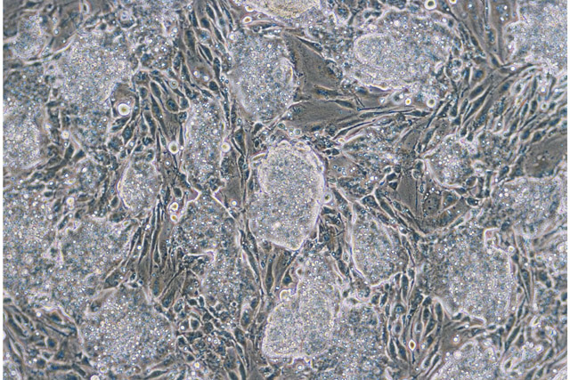
ES cells are grown on a layer of feeder cells, which are inactivated by irradiation or by the treatment with chemicals that prevent cell division.
ES cells require careful attention to be grown successfully or they can spontaneously differentiate and lose their ability to become all cell types within the body.
Undifferentiated ES cells form tight clusters of cells, called colonies, which have well defined boundaries when viewed under the light (phase contrast) microscope.
|
 |
| |
Top of Page |
| |
|
ES CELLS DIFFERENTIATED INTO CARDIOMYOCYTES
Without feeder cells or growth factors to maintain ES cells in an undifferentiated state, ES cells spontaneously differentiate into tissues.
For instance ES cells can form heart cells, called cardiomyocytes. These heart tissues are able to beat, as normal heart tissue.
However, differentiating ES cells are typically unable to form the true organs in culture, as they lack the signaling cascades and gradients which define the spatial matrix for the developing organ.
|
|
| |
Top of Page |
| |
|
|
|
Embryoid Body (EB)
Models |
Embryoid bodies are derived from ES cells, by growing ES without feeder cells. Without feeder cells ES cells will spontaneously differentiate into tissues of the body or can become simple embryo like structures.
Embryoid bodies offer a unique mechanism of studying development, in a simpler system, than the whole organism. EBs can be readily grown in tissue culture and can be influenced and directed down specific developmental paths by the addition of growth factors to the culture media.
The effect of genetic modifications can also be studied in EBs, by introducing genetic modifications into the ES cells from which the EBs are derived.
|
|
|
| |
Top of Page |
| |
|
ES CELLS FORM EMBRYOID BODIES (EBs)
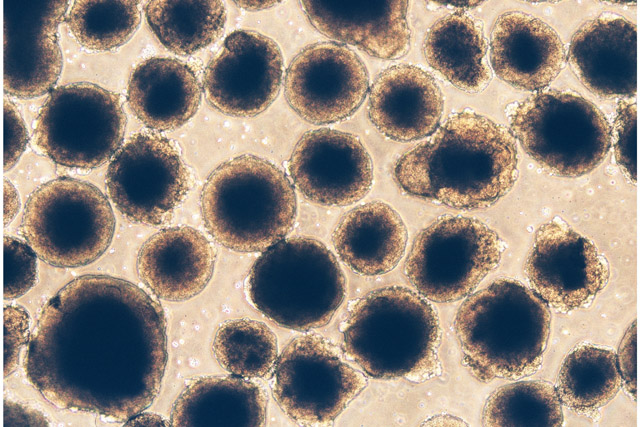
ES cells which are grown without feeder cells and which are prevented from attaching to the culture plate, can form embryo like structures, called embryoid bodies.
Embryoid bodies develop into rudimentary tissues and can be used as a model system to observe early tissue and organ development.
EBs are particularly useful in the study of blood vessel formation,
as all tissues within the body require the development of blood vessels. Therefore, all EBs will potentially have blood vessel formation, as they increase in size. |
 |
| |
Top of Page |
| |
|
MODELS OF BLOOD VESSEL FORMATION
An understanding blood vessel formation is important, as all tissues, including cancers (solid tumors), require a blood supply for growth, development and metastasis.
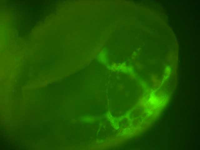
Most developing EBs develop endothelia cells, which are the initial cell type in developing blood vessels. Endothelial cells and developing blood vessels can be readily stained in whole or sectioned EBs.
In the panel to the right whole EBs have been stained with a fluorescent labeled antibody to an endothelia protein and the beginnings of blood vessels can be readily observed (bright green areas).
The EB blood vessel model can be very useful to identify genes involved in blood vessel formation, growth factors and drugs which promote or retard blood vessel formation.
Much interest is focused on identifying drugs which can inhibit blood vessel formation, as these agents may have important anti-tumor properties. Solid tumors must develop blood vessels in order to grow and survive. Under normal circumstances normal tissues in the adult only require blood vessel formation during wound healing. Therefore, a drug which inhibits blood vessel formation, may inhibit tumor development in adults and not have serious side effects.
|
 |
| |
Top of Page |
| |
|
|