|

|
|
|
| The Need For Stereology? |
I am interested in the collection of information and how this is used to build an accurate understanding of the biological world. In particular, I am interested in applied biological problems, such as the assessment and development of disease therapies.
Over the years it has become apparent that there is a strong need for appropriate methods to measure structures, in order to correctly and accurately understand the world.
A number of serious errors can be made when quantitating and assessing the real world. Benoit Mandebrot touched on these problems when he discussed measuring the length of the coastline using yard sticks of different dimensions. The shorter the yard stick, the longer the reported length of the coastline, compared to the results from the longer yard stick. Therefore, in the real world, length only has meaning when viewed in the context of the measuring device.
Other misconceptions and limitations in understanding the world are also highlighted by the issue of context. For example, when trying to understand brain function, it is important to measure not only the absolute number of neurons within the brain, but also the relative number of neurons to total cells, the complexity of the individual neurons and the relative complexity of neurons to the complexity of the brain structure itself.
Although brain damage can legitimately be caused by a decrease in the absolute number of neurons, changes in the proportion of neurons to supporting cells, loss of individual neuron's complexity or changes in the relative complexity of the neuron's to overall brain structure can also account for brain damage. Therefore, assessing brain damage solely based upon assessing the absolute number of neurons could easily give an inaccurate and incomplete understanding of brain function and the causes of brain damage.
|
| |
Top of Page |
| |
|
|
|
PROBLEMS WITH VISUALIZATION
A number of techniques are available to visualize the development or presence of structures within tissues, organs and the whole organism.
One such technique which allows for the visualization of developing blood vessels in whole EBs is confocal microscopy. In this technique, whole EBs can be optically sectioned and recorded using a scanning laser beam.
In the panel to the right a single EB, which has formed a blastocyst (embryo) like structure, has been stained to show endothelial cells (developing blood vessels). The location of the blood vessels within the whole EB was visualized using confocal microscopy.
The location of endothelia cells within the single EB can be seen clearly. However, to correctly understand blood vessel formation within EBs and the effects of genes or drugs on blood vessel formation within EBs, a number of questions must be addressed. For instance, how representative is any single EB compared to all EBs grown under the same growth conditions? What is the effect of the size, shape and complexity of the whole EB on the number of endothelial cells and the location of the endothelial cells?
Although confocal microscopy is a powerful method to visually demonstrate structures, the technique cannot quickly or readily quantitate structures within many samples. Another approach is necessary to quickly and efficiently quantitate the relative volume, length and complexity of blood vessels, compared to the varied and ever changing shapes of the embryoid body. |
|
| |
Top of Page |
| |
|
|
|
| Stereology Is Not! |
Stereology is a method of quantitatively assessing the relative number, volume and complexity of structures. The outputs of stereology are typically presented as tables and graphs. Therefore, stereological outputs are not as glamorous as the visual representations of structures derived from techniques like confocal microscopy; which beautifully present structures pictorially.
Stereology does not redraw structures from 2D sections, but does efficiently and quantitatively define the numbers of structures, their lengths, volumes and complexity. These assessments are vitally important to the correct understanding of the world, as the information derived from the more pictorial methods typically only represents a limited sample area and frequently is not a true quantitative representation of the 3D structure scanned. |
| |
Top of Page |
| |
|
| What Is Stereology? |
Stereology is a process for measuring structures, within a reference space and uses a combination of statistics and geometry to obtain and formulate an unbiased numerical assessment of the original 3D structure.
Information about 3D structures is typically obtained from 2D sections of the structure. Even within modern imaging techniques, such as MRI and CAT scans, 3D images are assembled from a large number of 2D sections.
2D sections must typically be acquired, as it is frequently impossible to collect 3D information during a single time point. This presents a significant problem; how do you piece 2D information together to accurately represent 3D structures?
Although stereological approaches compliment 3D visualization techniques, stereology has several important advantages over them. Stereology represents an unbiased and efficient approach to understanding structures, which doesn't require huge computational capabilities. Using stereological techniques, it is possible for a very small number of unbiased data points to correctly and accurately describe any structure. |
| |
Top of Page |
| |
|
| How it works |
In order to quantitatively assess structures within tissues, organs or the whole organism, 2D sections must be acquired, the structures of interest identified (typically through staining) and the number of those structures of interest appropriately counted within the sections.
To correctly piece together this information and understand the structures of interest within the reference space, the proceeding steps must all be carried out impartially, using statistically sound procedures. Therefore, how the structures are embedded, sectioned and counted, must be carefully considered and carried out in such a way as to avoid introducing bias.
Once sections are stained for the structures of interest, a counting grid is overlaid on top of photographs of the stained sections. The number of times that structures intersect with the counting grid is determined. By varying the nature and shape of the counting grid, assessment of absolute number of items, lengths, volumes or complexity of items within the reference space can be achieved. |
| |
Top of Page |
| |
|
| Quantitation of Blood Vessel Production In EBs |
The development of blood vessels within EBs can be influenced in a number of ways; genetic modifications can be introduced into the ES cells from which the EBs are derived or EBs can be grown in growth media supplemented with chemicals which affects blood vessel formation and development.
Blood vessel formation can be monitored within whole EBs, by staining with dyes specific for blood vessel formation and recording the results using microscopic analysis, such as confocal microscopy.
Although this approach allows for the visual inspection of EBs and blood vessel formation, this approach doesn't convey how blood vessel formation varies between EBs grown under the same conditions and does not quantitatively assess the amount of blood vessel formation.
Embedding EBs grown under the same conditions, sectioning embedded EBs followed by the counting of structures using stereologically appropriate procedures allows for the efficient and accurate assessment of the influence of factors on blood vessel formation within EBs.
|
| |
Top of Page |
| |
|
|
|
|
 |
 |
 |
| |
|
|
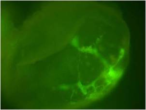
Media Only
Blood vessels develop in whole growing EBs when cultured in
regular growth media. |
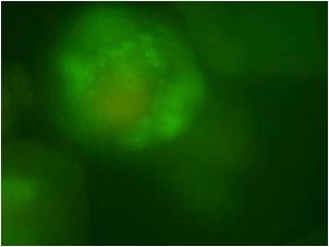
Blood Vessel Inhibitors
Differentiation inhibitors retard both
whole EB development and alter
blood vessel network formation. |
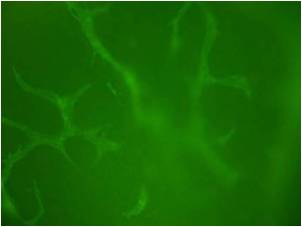
Blood Vessel Promoters
Blood vessel promoters appear to generate larger EBs, with increased blood vessel formation. |
| |
|
|
| |
|
Top of Page |
| |
|
|
|
|
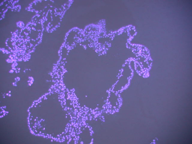
SECTIONS STAINED FOR EB STRUCTURE
A large number of EBs grown under specific growth conditions can be readily embedded and sectioned.
The structure of EBs can be visualized in sections and their area, volume and complexity can be assessed using stereological techniques.
A sectioned EB can be seen stained blue, in the panel to the right. The stained EB shows a large hollow central cavity, similar to the structure of the developing blastocyst in the normal embryo.
|
 |
| |
Top of Page |
| |
|
SECTIONS STAINED FOR ENDOTHELIAL STRUCTURES
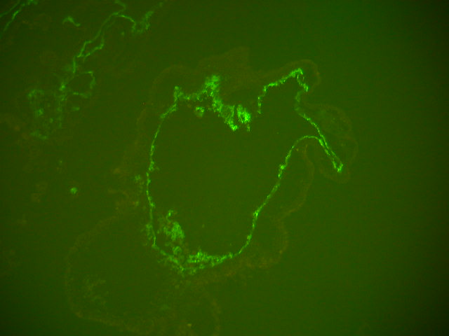
A subsequent section of the EB shown above, was stained for blood vessel formation and is shown in the panel to the right.
Blood vessel formation can be discerned in growing EB, although the 3D pattern of the developing vessels cannot be determined in a single section.
From the section, it can be noted that blood vessel formation appears to occur within the center of the developing EB. Possibly as the exterior of the EB can derive sufficient oxygen and nutrients from the culture media. This may reduce the need for vessel formation on the outside of the EB.
|
 |
| |
Top of Page |
| |
|
| |
|
EB VOLUME
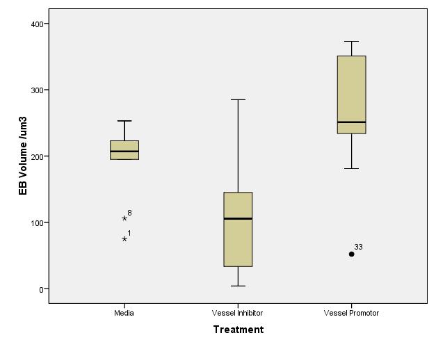
The boxplot to the right shows the volume of EBs grown in media, supplemented with various drugs.
From the boxplot, it can be noted that EBs grown in media only tend to have a consistent size, with a lower median size than EBs grown in the presence of drugs which promote blood vessel formation.
EBs grown in media supplemented with blood vessel inhibitors, appear significantly smaller than those grown in media only, but have a very wide range of sizes.
EBs grown in media supplemented with blood vessel promoters have a varied size and are significantly larger than EBs grown in media only. |
 |
| |
Top of Page |
| |
|
BLOOD VESSEL VOLUME
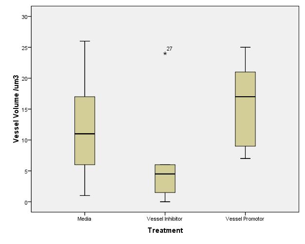
The boxplot to the right shows the volume of blood vessels within EBs grown in media, supplemented with various drugs.
EBs grown in media only, have a wide range of blood vessel volumes, with a median vessel volume that is significantly less than that of EBs grown in blood vessel promoters.
Those grown in media supplemented with blood vessel inhibitors, have a narrow range of blood vessel volumes and a significantly lower median vessel volume than EBs grown in media only.
EBs grown in media supplemented with blood vessel promoters, have a wide range of vessel volumes with an significantly higher median vessel volume than EBs grown in media only. |
 |
| |
Top of Page |
| |
|
BLOOD VESSEL VOLUME FRACTION
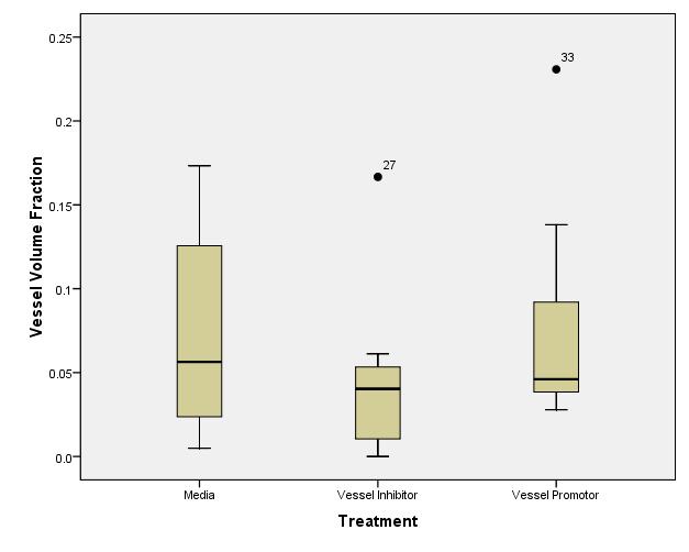
The boxplot to the right shows the fractional volume of blood vessels to total EB volume, within EBs grown in media supplemented with various drugs.
It can be noted that EBs grown in media only have a wide range of fractional blood vessel volumes to total EB volume, with a median blood vessel volume fraction of about 6%.
EBs grown in media supplemented with blood vessel inhibitors have a narrow range of blood vessel volume fractions, with a lower median blood vessel volume fraction, compared to media only, of about 4%.
EBs grown in media supplemented with blood vessel promoters have a modest range of blood vessel volume fractions, compared to EBs grown in media only, with a median blood vessel volume fraction of about 5%. |
 |
| |
Top of Page |
| |
|
|
|
| Understanding Blood Vessel Formation in EBs |
As stereological techniques provide quantitative data about structures, the information from these analyses can be important in the understanding of biological processes. For instance, the results of the combined analysis of EB development in the presence of blood vessel inhibitors and promoters provide information about the mechanisms of EB growth and development.
The treatment of EBs with blood vessel inhibitors significantly reduces both the median volume of blood vessels and EBs. In addition, blood vessel inhibitors reduce the dispersion of blood vessel volume and blood vessel volume fraction in EBs. Taken together, these results indicate that blood vessel inhibitors are highly effective at preventing the development of blood vessels within EBs which directly leads to a decrease in the size attained by EBs.
Treatment of EBs with blood vessel promoters significantly increases the median blood vessel volume and median size of the EBs, but does not significantly affect the median blood vessel volume fraction. Taken together, these results indicate that EBs require a minimum blood vessel volume fraction to grow and acquire overall volume. Interestingly, the increase in both the dispersion of EB volume and blood vessel volumes support the idea that blood vessel formation may be a limiting factor in the growth and development of EBs in culture.
|
| |
Top of Page |
| |
|
|